Keterangan
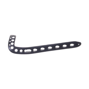
Sistem Pelat Osteotomi HTO
Q&A
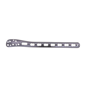
Q1: What are the primary functions and biomechanical differences between HTO Osteotomy Plate System? A1:Lateral Distal Femoral Osteotomy Plate: Used for medial closing wedge femoral osteotomy to correct valgus deformity; must withstand high femoral loads.
Medial Distal Femoral Osteotomy Plate: Used for lateral closing wedge or medial opening wedge to correct varus deformity; lower profile due to medial soft tissues.
Medial High Tibial Osteotomy Plate: Opening wedge design for varus correction; relies on intact lateral hinge and often incorporates a spacer.
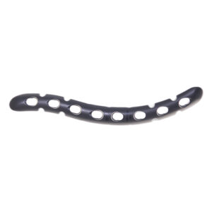
Lateral High Tibial Osteotomy Plate: Closing wedge design; provides inherent stability from bone-to-bone contact but removes bone.
Q2. How does the biomechanical performance differ between medial and lateral HTO plates?
A2: Medial HTO plates function as tension bands withstanding tensile forces, while lateral HTO plates serve as compression devices. Medial designs require stronger buttress support against varus collapse, whereas lateral designs focus on maintaining compression across the osteotomy site.
Q3:What are the key surgical techniques for preventing non-union in HTO procedures?
A3: Critical techniques include preserving a 5-10mm intact lateral hinge, using adequate bone graft for openings >10mm, maintaining stable fixation with locking screws, and ensuring proper plate contouring to match bone anatomy.
Q4:How do patient factors influence plate selection in HTO surgery?
A4: Key factors include bone quality (locking screws for osteoporosis), BMI (stronger constructs for obese patients), age (consider biological healing potential), and activity level (optimize stability for active patients).
Q5: How does wedge size affect fixation strategy in opening vs closing wedge osteotomies?
A5: Larger wedges (>12mm) require structural bone graft, additional anti-glide plating, and extended protection periods. Closing wedges provide inherent stability but may cause leg length discrepancy.
Q6: What is the typical weight-bearing progression for each osteotomy type?
A6:Medial HTO: 6-8 weeks non-weight-bearing, gradual progression to full weight-bearing by 12 weeks.
Lateral HTO: Earlier weight-bearing possible (4-6 weeks) due to inherent stability.
Distal Femur osteotomy: Longer protection period (8-12 weeks non-weight-bearing) due to higher forces.
Q7: What are the key radiographic parameters for assessing osteotomy healing?
A7:Tibial Osteotomies: Healing of medial hinge, maintenance of correction angle, trabeculation across osteotomy site.
Femoral Osteotomies: Cortical continuity, callus formation at osteotomy gaps, hardware position.
All Procedures: Mechanical axis alignment on standing full-length films.
Q8: When is combined femoral and tibial osteotomy indicated?
A8: Combined procedures are needed for corrections >15°, significant joint line obliquity (>10°), or double-level deformities. They require careful sequencing and potentially hybrid fixation strategies.