What is Osteogenesis Imperfecta (HEI)?
Osteogenesis imperfecta (HEI) is a genetic or heritable disease in which bones fracture (break) easily, often with no obvious cause or minimal injury. OI is also known as brittle bone disease, and the symptoms can range from mild with only a few fractures to severe with many medical complications.
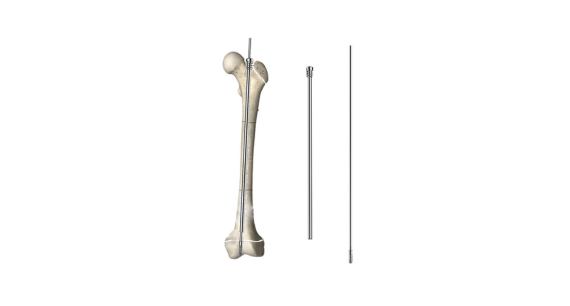
Telescopic intramedullary nail is a self extending rod designed for patients suffering from Osteogenesis Imperfecta (HEI), displasia rangka dan kecacatan tulang lain. Created to prevent or stabilize fractures, or correct deformity of long bones whilst growth occurs.
The design of the rod includes a female component (which is attached to the proximal epiphysis) and a male component (which is attached at the distal epiphysis).They extend together,without hindering bone elongation.

Operation Technique of Pediatric Telescopic Nail
1.Locate the osteotomy point (CORA point), predict the body surface position of the osteotomy point, place markers, and C-back fluoroscopy.
2.Reaming the marrow
Choose 0.2-0.3 mm greater than the required mother needle hollow drill, insert the needle, by the end of bone cutting proximally or distal pulp, and proximal enlarge pulp can be conveniently from the great trochanter inside out, Find the needle in the proximal point, proximal cut soft tissues such as skin.
3.After insert core needle enlarge pulp, pull out the needle, from the cut after proximal bone of extremities retrograde insertion, suitable core needle is inserted into the hollow screw driver.
4.Implant the core needle into the distal epiphysis. C-arm perspective anteroposterior and lateral view: the threaded part of the core needle is implanted into the distal epiphysis, not beyond the articular surface, and is in the anteroposterior and lateral center.
5.Implant the core needle into the distal epiphysis. C-arm perspective anteroposterior and lateral view: the threaded part of the core needle is implanted into the distal epiphysis, not beyond the articular surface, and is in the anteroposterior and lateral center
6.Keep the tail of the core needle 5-10mm higher than the female needle (reserve growth space), cut off the excess core needle.
7.The probe probes the tail of the core needle to ensure that the tail is smooth and does not affect the relative sliding of the core needle and the female needle during bone growth.
X-Ray


About CareFix
CareFix is a professional national high-tech enterprise engaged in orthopedic medical equipment design, pengeluaran, sales and service. Founded in 2009, we’ve devoted to pediatric implants,kaki & Implan buku lali,external fixation and spinal implants. Welcome to visit to our factory,it’s about half an hour from Shanghai Hongqiao Airport.